Algae Cell Counting Is Broken: Why Researchers Are Switching to Automated Analysis
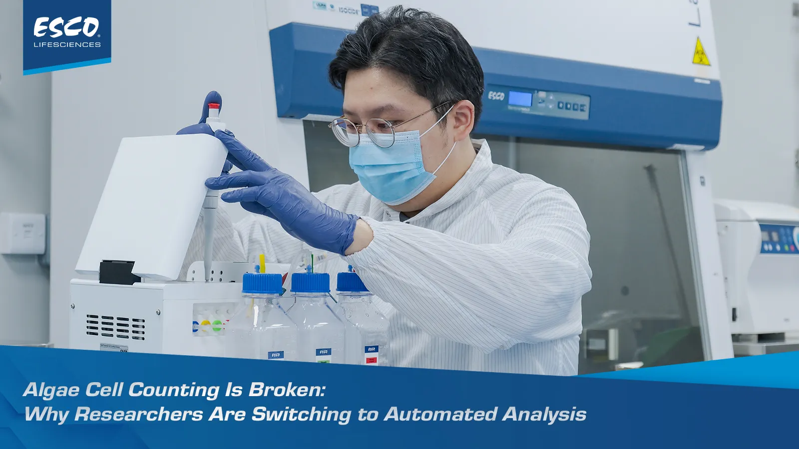
In algae research and industrial cultivation, everything downstream — growth curve tracking, harvest timing, strain screening, process optimization — depends on one number: how many cells are actually in your sample. Yet accurate, rapid algae cell counting remains one of the most persistent bottlenecks in the workflow. Most labs are stuck choosing between methods that are fast but unreliable, or accurate but painfully slow.
This trade-off isn't a training problem or a technique problem. It's a limitation baked into the traditional tools themselves. Below, we break down exactly where each conventional method falls short — and how a new generation of automated cell counting technology closes the gap.
The Problem With Traditional Algae Counting Methods
Algae counting has historically relied on three approaches, each with a well-documented weak point.
1. Microscope Counting (Hemocytometer): Accurate in Theory, Costly in Practice
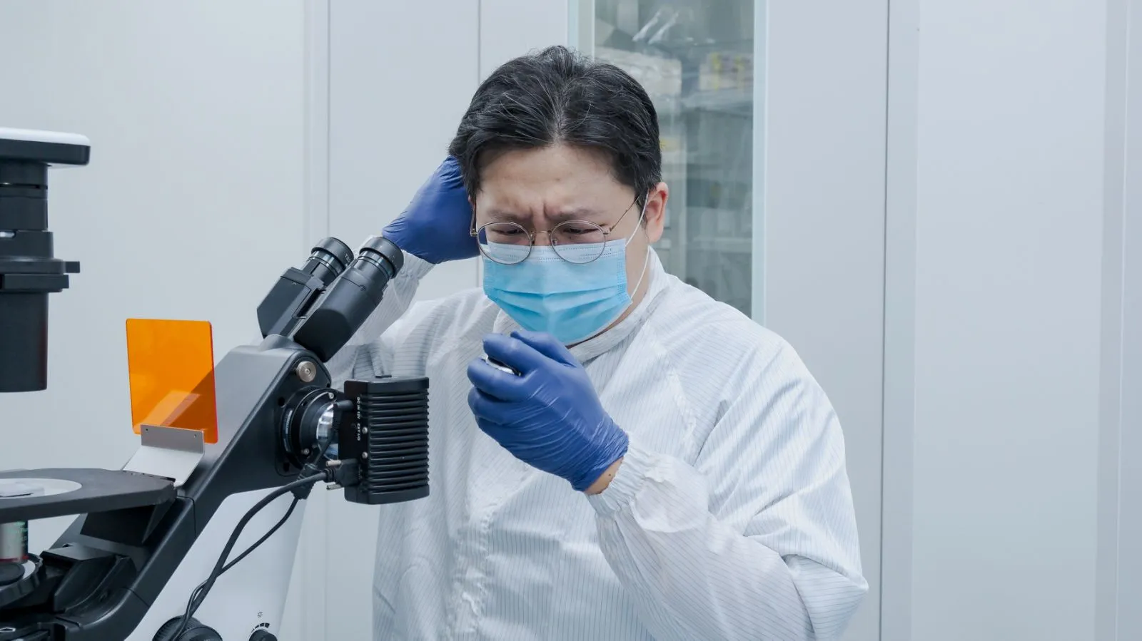
Direct microscope counting is the classic, go-to method for algae cell counting. It's intuitive: place the sample under the lens, count what's in the field of view, extrapolate.
The pain point: it's slow and labor-intensive, and results swing based on the algae's own physiological state, suspended particles in the water, and — perhaps most critically — the individual operator. Two technicians counting the same sample can land on two different numbers.
2. Turbidity (OD Value) Measurement: Fast, But Not Actually Counting Cells
The turbidity method estimates algae concentration indirectly, by measuring how much light the culture medium absorbs. It's simple and supports high-throughput testing.
The pain point: a standard curve has to be re-drawn before every test run, which adds friction to an otherwise quick method. More fundamentally, turbidity never counts a single cell — it infers a number from absorbance, which means shifts in cell size, shape, or pigmentation can quietly distort the result.

3. Image-Based Photography Counting: Sees Part of the Picture, Not All of It
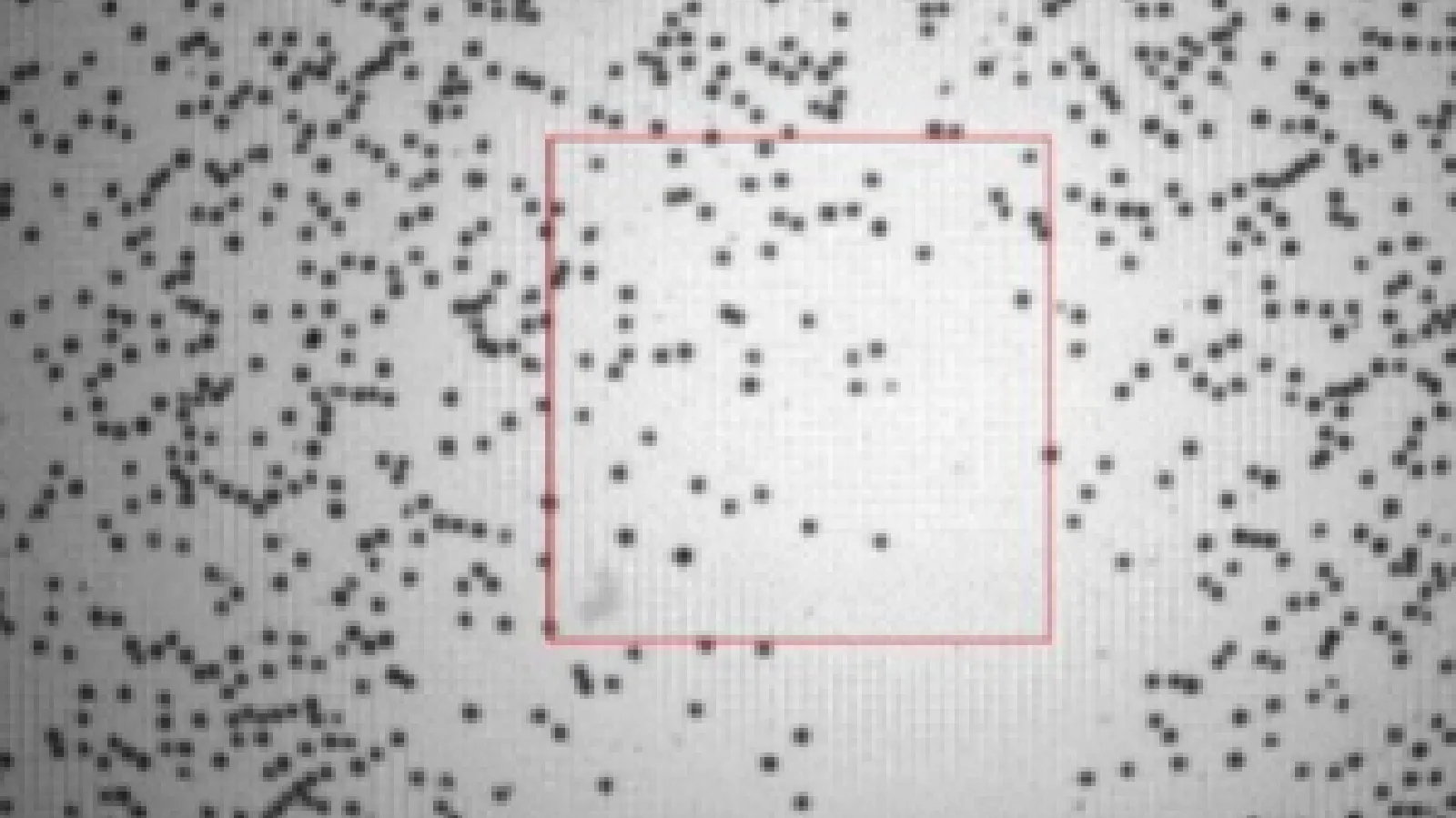
Image-based counters photograph a sample and use algorithms to automatically identify and count cells. It's fast and simple to run.
The pain point: these systems typically image only a portion of the sample chamber, not the full volume — so the count is a projection, not a direct measurement. Accuracy also depends heavily on the algorithm correctly recognizing cell shape and type, which becomes unreliable across algae's wide morphological diversity.
The Solution: Esco ACC Automated Cell Counting Analyzer
Esco Lifesciences developed the ACC series Automated Cell Counting Analyzer specifically to remove this speed-versus-accuracy trade-off. Instead of choosing between the strengths of microscopy, turbidity, or imaging, the ACC combines Coulter principle technology with intelligent image analysis — a full-sample, direct-measurement approach purpose-built for algae's counting challenges.
Here's how each core advantage of the ACC directly answers a limitation above.

Core Advantage 1: A Complete-Sample Count, Not a Partial Field of View
The ACC uses the Coulter principle to physically measure every cell that passes through it, rather than sampling one field of view (as with microscopy) or one imaged region (as with photography-based counters).
- Every single cell in the sample is counted — no sampling bias
- Coefficient of variation (CV) stays below 5%, for highly reproducible results
- The Coulter principle is a recognized standard reference method, measuring actual cell volume rather than a projected image area
This solves the operator-dependent variability of microscope counting and the partial-view sampling gap in image-based methods.
Core Advantage 2: Precise Counting Regardless of Cell Shape
Algae are morphologically diverse — Chlorella is spherical, Microcystis forms clumps, and diatoms vary in geometric shape. Image-recognition algorithms often struggle to keep up.
The ACC detects cells by changes in electrical resistance, not by visual shape recognition — so spherical, ellipsoidal, or conical cells are all counted with the same precision. Its intelligent algorithm also automatically separates clumped cells, resolving a common failure point for aggregated samples.
This solves the shape- and algorithm-dependent accuracy problems inherent to image-based counting.
Core Advantage 3: Real-Time Detection, No Settling Delays
Algae cells settle when left standing, which skews sample uniformity and throws off counting accuracy in static methods.
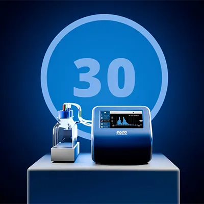
The ACC's microfluidic dynamic detection technology counts cells in real time as they flow — with no waiting for settling equilibrium and no accuracy loss from stratification. Results are ready in under 30 seconds from sample loading.
This solves the time-consuming setup of microscope counting and the standard-curve overhead of turbidity measurement.
Core Advantage 4: Reusable Chip Design, Lower Cost Per Test
Most image-based counters require disposable counting plates or chips, which drives up long-term operating costs.
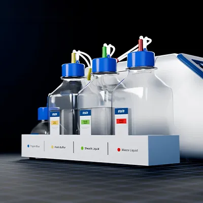
The ACC uses a reusable microfluidic chip, cutting long-term consumable costs by more than 50%. It's also compatible with direct dilution and loading of culture medium samples, simplifying sample preparation and maintenance.
This solves the recurring consumable cost and prep complexity of disposable-plate imaging systems.
The Data: Field-Tested Performance
Independent field testing on algae samples validated the ACC across three critical performance dimensions:
| Performance Metric | ACC Result | Why It Matters |
|---|---|---|
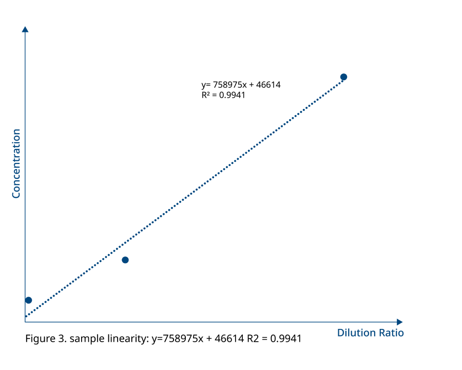
| Linearity | Detected concentrations closely matched theoretical dilution ratios across serial dilutions | Confirms precise, dependable quantification across a wide concentration range — not just at one convenient dilution point |
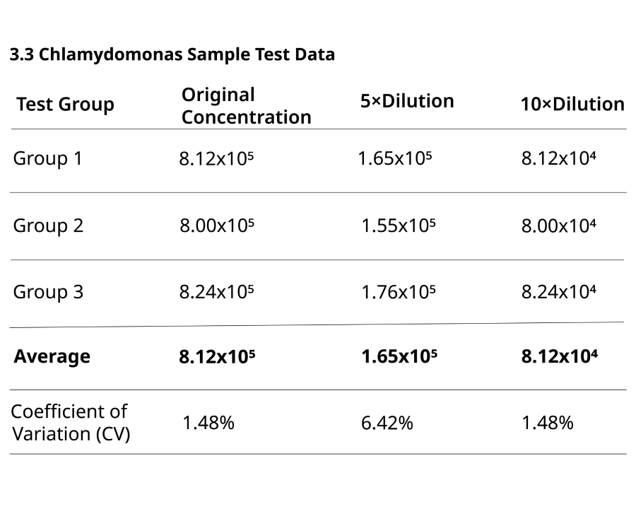
| Reproducibility (CV) | 1.48% – 7.46% | Far below the >12% CV typical of manual counting, meaning results are consistent between repeat tests and repeat operators |
| Detection Range | 5.0 × 10⁴ to 2.0 × 10⁷ cells/mL | Covers algae culture from low-density starts through the logarithmic growth phase, without switching instruments or methods |
Taken together, this data shows an instrument that stays accurate whether a sample is dilute or dense, and stays consistent whether a technician runs it once or a hundred times.
The Bottom Line
Every advance in algae research and industrial-scale cultivation ultimately rests on trustworthy cell density data. The Esco ACC Automated Cell Counting Analyzer was engineered to remove the traditional trade-off between speed and accuracy — pairing Coulter principle physical measurement with intelligent image analysis to deliver full-sample counts, shape-independent precision, real-time results, and a lower cost per test.
Ready to see how the ACC performs on your own algae samples? Contact Esco Lifesciences to arrange a demonstration or request detailed application data for your strain and workflow.